Conditions
and
treatments
From macular degeneration to diabetic retinopathy, timely treatments illuminate paths to sight, safeguarding our vision's vibrant tapestry.
-
Macular disease refers to any condition that affects the macula, the central part of the retina responsible for sharp, central vision. This can include conditions such as macular degeneration, macular edema, macular hole, and macular pucker. Symptoms of macular disease may include blurred or distorted central vision, straight lines appearing wavy or bent, or a dark spot in the center of your vision.
Treatment for macular disease depends on the specific condition and may include medications, laser therapy, injections of anti-VEGF medications, or surgical procedures such as vitrectomy. Early detection and treatment are crucial for preserving vision and preventing permanent vision loss.
-
Macular degeneration is a common eye condition that affects the macula, a small area in the center of the retina responsible for sharp, central vision. There are two types of macular degeneration: dry and wet. Dry macular degeneration occurs when the cells in the macula break down gradually, leading to blurred or distorted central vision. Wet macular degeneration is more severe and involves the growth of abnormal blood vessels under the macula, which can leak fluid and cause rapid vision loss.
Treatment for macular degeneration depends on the type and severity of the condition. In the early stages, lifestyle changes such as quitting smoking, eating a healthy diet rich in antioxidants and omega-3 fatty acids, and protecting your eyes from UV light may help slow the progression of the disease. In more advanced cases, treatments such as injections of anti-VEGF medications or photodynamic therapy may be recommended to reduce abnormal blood vessel growth and preserve vision.
-
Hypertensive retinopathy is damage to the retina caused by high blood pressure. Chronic hypertension can cause narrowing of the blood vessels in the retina, leading to decreased blood flow and oxygen delivery to the retinal tissue.
Symptoms of hypertensive retinopathy may include vision changes, such as blurred or decreased vision, and in severe cases, swelling of the optic nerve head (papilledema). Management of hypertensive retinopathy involves controlling blood pressure through lifestyle modifications and medications to prevent further damage to the retina and preserve vision.
-
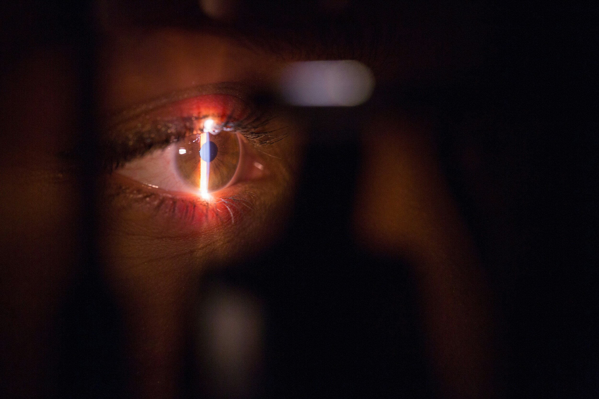
A diabetic eye exam is a comprehensive eye examination performed to assess the health of the eyes in people with diabetes. Diabetes can cause damage to the blood vessels in the retina, leading to a condition called diabetic retinopathy, which can cause vision loss if left untreated. During a diabetic eye exam, the eye doctor will dilate the pupils and examine the retina for signs of diabetic retinopathy, including swelling, leaking blood vessels, or abnormal growth of blood vessels.
Early detection and treatment of diabetic retinopathy are crucial for preserving vision and preventing complications. People with diabetes should have a dilated eye exam at least once a year, or more frequently if recommended by their eye doctor.
-
Diabetic retinopathy is a complication of diabetes that affects the blood vessels in the retina. High blood sugar levels can damage the blood vessels, causing them to leak fluid or bleed into the retina, leading to vision loss. There are two main stages of diabetic retinopathy: non-proliferative diabetic retinopathy (NPDR) and proliferative diabetic retinopathy (PDR). NPDR is the early stage, where small blood vessels in the retina leak fluid or blood. PDR is more advanced and involves the growth of new, abnormal blood vessels on the retina, which can bleed and cause scarring.
Treatment for diabetic retinopathy depends on the stage and severity of the condition. In the early stages, close monitoring of blood sugar levels and blood pressure, along with regular eye exams, may be sufficient to prevent vision loss. In more advanced cases, treatments such as laser therapy, injections of anti-VEGF medications, or vitrectomy surgery may be necessary to preserve vision and prevent further damage to the retina.
-
Intraocular injections involve administering medication directly into the eye. These injections are commonly used to treat various retinal conditions such as macular degeneration, diabetic retinopathy, and retinal vein occlusion. The medications injected into the eye may include anti-VEGF drugs, steroids, or other agents aimed at reducing inflammation, preventing abnormal blood vessel growth, or promoting healing.
While intraocular injections can be effective in managing certain retinal diseases, they do carry some risks, including infection and increased eye pressure. Therefore, these injections are typically performed by trained ophthalmologists in a controlled clinical setting.
-
A retinal tear occurs when the thin lining at the back of your eye, called the retina, gets a rip or a break. This can happen due to aging, trauma, or other eye conditions. Retinal tears are serious because they can lead to retinal detachment, where the retina pulls away from the back of the eye, causing vision loss.
Symptoms of a retinal tear may include sudden flashes of light, floaters (tiny specks or threads in your vision), or a curtain-like shadow in your peripheral vision. If you experience any of these symptoms, it's essential to see an eye doctor right away.
Treatment for a retinal tear often involves sealing the tear to prevent fluid from getting behind the retina and causing detachment. This is typically done through a procedure called laser photocoagulation or cryopexy, where the tear is sealed using a laser or freezing technique. Early detection and treatment of retinal tears are crucial for preventing vision loss.
-
A retinal detachment occurs when the retina pulls away from the back of the eye, causing vision loss. This can happen due to trauma, aging, or underlying eye conditions such as lattice degeneration or diabetic retinopathy. Symptoms of a retinal detachment may include sudden flashes of light, floaters (dark spots or lines in your vision), or a curtain-like shadow in your peripheral vision.
A retinal detachment is a medical emergency that requires prompt treatment to prevent permanent vision loss. Surgery is usually necessary to repair a retinal detachment and reattach the retina to the back of the eye. There are several surgical techniques used to treat retinal detachments, including scleral buckling, vitrectomy, and pneumatic retinopexy. The choice of treatment depends on the location and severity of the detachment.
-
A macular hole is a small hole that develops in the macula, the central part of the retina responsible for sharp, central vision. A macular pucker, also known as epiretinal membrane, is a thin layer of scar tissue that forms on the surface of the macula, distorting vision. Both conditions can cause blurred or distorted central vision.
Treatment for macular holes and puckers may involve vitrectomy surgery, where the vitreous gel inside the eye is removed, and the macular hole or pucker is repaired. During the procedure, the surgeon may use special instruments to peel away the scar tissue or close the hole with a gas bubble. Recovery from macular hole and pucker surgery can take several weeks, and vision may continue to improve gradually over time.
-
Scleral buckling is a surgical procedure used to repair retinal detachments. During the procedure, a silicone band or sponge is placed around the outside of the eye to indent or "buckle" the sclera, the white outer layer of the eye. This indentation helps to bring the detached retina back into its proper position against the back wall of the eye.
Scleral buckling surgery is often performed in conjunction with other procedures such as vitrectomy, which involves removing the gel-like substance inside the eye (the vitreous). Together, these techniques help to reattach the retina and restore vision in cases of retinal detachment.
-
A vitrectomy is a surgical procedure used to remove the vitreous gel from the middle of the eye. This procedure is commonly performed to treat conditions such as retinal detachment, macular hole, or vitreous hemorrhage.
During a vitrectomy, the surgeon makes small incisions in the eye and uses tiny instruments to remove the vitreous gel and any scar tissue or debris that may be affecting vision. After the vitreous is removed, it may be replaced with a clear solution or gas bubble to help support the retina while it heals.
Vitrectomy surgery can help restore or improve vision in many cases, but it does carry risks, including infection, bleeding, and retinal detachment. Your surgeon will discuss the potential benefits and risks of vitrectomy surgery with you and determine if it's the right option for your specific eye condition.
-
Panretinal photocoagulation (PRP) is a laser treatment used to treat proliferative diabetic retinopathy. During the procedure, laser burns are applied to the peripheral areas of the retina to reduce abnormal blood vessel growth and prevent bleeding and retinal detachment.
PRP treatment can help preserve vision and reduce the risk of severe complications in patients with diabetic retinopathy. However, it may cause temporary vision changes and discomfort immediately following the procedure.